The Neuroimage Analysis Laboratory
Accelerating biology through quantitative analysis of images and data mining
Core overview
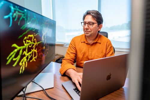
Imaging is a primary method to capture complex biological processes at multiple scales that range from single molecules to whole-body. We use many modalities in our laboratory, including light and electron microscopy, ultrasound, PET and MRI and regular cameras. It is the focus of the Neuroimage Analysis Laboratory (NIAL) to leverage advanced image processing and analysis methods to turn the images into quantitative knowledge. Our services include:
- Project consultation and planning
- Image processing (deconvolution, deskew, denoising, etc.)
- Animal behavior analysis
- Image registration
- Image segmentation, classification and object tracking
- Data mining using machine-learning
- Image data visualization
Research summary
Recent advancements in the field of computer vision have revolutionized how images are analyzed. The Neuroimage Analysis Laboratory (NIAL) aims to utilize these advancements and develop novel image- and data-analysis pipelines in close collaboration with wet-lab researchers. NIAL maintains and provides easy access to a range of high-performance servers and workstations to support researchers with their image and data analysis computational needs.
Key Equipment List
Shared Linux Server:
- Ultra94: Intel Xeon 96 CPU cores 1.5TB RAM
- DNB1: AMD EPYC 128 CPU cores 2TB RAM, 2 x Nvidia RTX A6000 GPU
- DNB2: AMD EPYC 128 CPU cores 1TB RAM, 2 x Nvidia RTX A6000 GPU
Dedicated Linux Servers:
- GPX10: Intel Xeon 20 CPU cores 512GB RAM - 10 x Nvidia 1080 ti GPU
- OMERO: Intel Xeon 96 CPU cores 768GB RAM - 1 x Nvidia 2080 ti GPU
Software Available on Linux Servers:
- Remote Desktop: NoMachine, FastX
- Image Analysis: Fiji, Ilastik, CellProfiler, Python, Matlab, 3DSlicer, Paraview
- Data Analysis: RStudio, Python, 10X Genomics
Shared Windows Workstations:
- Workstation #1: Intel i9 18 CPU cores 128GB RAM - 1x Nvidia 2080 ti GPU
- Workstation #2: Intel i9 10 CPU cores 64GB RAM - 1x Nvidia 1080 ti GPU
- Workstation #3: Intel Xeon 36 CPU cores 512GB RAM - 1x Nvidia 1080 ti GPU
Software Available on Windows Workstations:
- Image Visualization: Arivis 4D, SlideBook, ImageScope
- Image Analysis: Fiji, CellProfiler, Microvolution, RStudio, Matlab
About the director
Dr. Abbas Shirinifard completed his PhD in Physics at Indiana University in 2011, under supervision of Professor James Glazier, focused on angiogenesis in cancer and choroidal neovascularization. He then pursued a post-doc with Dr. Glazier studying the effects of arsenic on intersegmental vascular development in zebrafish embryos before joining St. Jude in 2012. In addition to his diverse experience in physics-based approaches of studying biological systems, he has hands-on experience in a wide range of in vivo and in vitro methods including light microscopy, contrast-enhanced ultrasound, xenografts, IHC and histology. As the core director, his multi-disciplinary background uniquely positions him to effectively collaborate with DNB labs, overcoming language barriers that often exist between wet-lab and computational scientists.

The Team
Benjamin Lansdell, PhD
Bioinformatics Research Scientist