St. Jude Family of Websites
Explore our cutting edge research, world-class patient care, career opportunities and more.
St. Jude Children's Research Hospital Home

- Fundraising
St. Jude Family of Websites
Explore our cutting edge research, world-class patient care, career opportunities and more.
St. Jude Children's Research Hospital Home
- Fundraising
Neuroimaging Laboratory
Accelerating biology through the application and development of advanced imaging methods
Overview
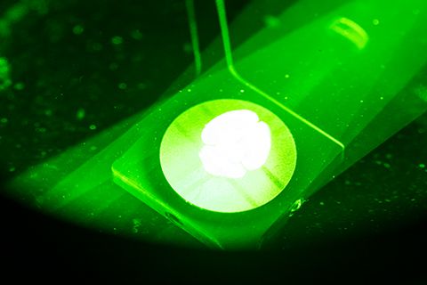
As the complexity of biological inquiry has increased, so too have the methods of study. Light microscopy is a hallmark technique that is well-poised to explore a variety of living biological systems, and recent advances in imaging technology expand our understanding like never before. These advanced imaging methods have traditionally been challenging to implement without domain expertise in optics and its interface with biology — skill sets not commonly present in a laboratory setting.
It is the focus of the Neuroimaging Laboratory (NIML) to expand access to and competency in advanced imaging methods at the institute. We accomplish this goal through the development and implementation of new methods with external collaborators in the private and academic sectors and through a robust training program that empowers biologists to fully leverage the latest methods.
Impact
The vast majority of scientific projects at St. Jude utilize light microscopy. This increasing competency serves as a catalyst to advance the core mission of understanding the processes that drive catastrophic pediatric diseases. Research in the Neuroimaging Laboratory (NIML) is currently focused on forging and maintaining key external collaborations to design and build custom microscopes that address critical needs in the department of Developmental Neurobiology.

Currently, the group is working on an adaptive optics enabled microscope (MOSAIC) that will allow researchers to visualize biological processes in deeper tissue layers by overcoming the optical barriers that make current methodologies difficult to apply. In parallel, the NIML is also focused on the continued development of training protocols for our instrumentation to ensure that the instruments we build and administer are used appropriately and to their fullest potential.
Equipment
Advanced Microscopy:
- Multi-modal optical scope with adaptive optics correction (MOSAIC)
- Two 3i Lattice Light sheet microscopes
- Miltenyi Ultramicroscope II Gaussian beam light sheet microscope
General Microscopy:
- Keyence BZ-X700 series with optical sectioning and incubation chamber
- Keyence BZ-X800
- Zeiss AxioImager with Apotome optical sectioning
- Zeiss SteREO Discovery.V12 stereomicroscope with fluorescence capability
Fabrication/Engineering capability:
- Harrick Plasma cleaner
- FormLabs Form2 SLA 3D printer
- Custom part design and prototyping upon request
Image Visualization:
- Arivis Vision4D
- Arivis InViewR VR visualization
- Paraview
- Blender
About the director
Daniel Stabley, PhD, grew up in the Metro Detroit area and attended Wayne State University where he obtained an honors college BS in Chemistry with an emphasis in Biochemistry. He then attended Emory University where he obtained his PhD under the mentorship of Khalid Salaita while working on novel biosensor development and microscopy methods. Stabley then completed a postdoctoral fellowship in the lab of David Solecki, PhD, at St. Jude where he studied neuronal cytoskeletal dynamics and force exertion during cerebellar development. In 2016 Stabley started the Neuroimaging Laboratory where he continues to pursue his passion for advanced microscopy and mentors users to equip the next generation of biologists at St. Jude and beyond with a firm foundation in advanced microscopy methods.

The team
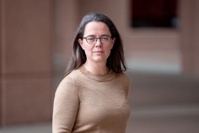
- Sharon King, PhD
- Senior Scientist
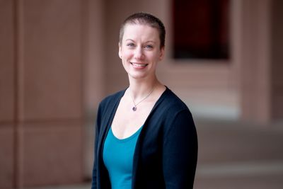
- Becky Petersen, PhD
- Lead Researcher
Memphis, TN, 38105-3678 USA GET DIRECTIONS