St. Jude Family of Websites
Explore our cutting edge research, world-class patient care, career opportunities and more.
St. Jude Children's Research Hospital Home

- Fundraising
St. Jude Family of Websites
Explore our cutting edge research, world-class patient care, career opportunities and more.
St. Jude Children's Research Hospital Home

- Fundraising
Asim Bag Lab
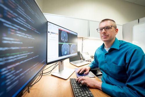
Harnessing multiparametric imaging and AI-driven analysis to characterize CNS tumors, predict tumor types and subtypes and assess therapy-induced damage to normal brain tissue.
About the Bag Lab
Our lab specializes in advancing the field of pediatric neuro-oncology through innovative applications of multiparametric imaging techniques. Since multiparametric imaging combines multiple imaging methods, these techniques allow us to precisely characterize various central nervous system (CNS) tumors. By using multimodal data, we are able to conduct comprehensive analyses and classification of tumor types and subtypes. We simultaneously use artificial intelligence (AI)-based methods to enhance diagnostic and predictive accuracy and efficiency. We apply an array of imaging techniques to perform quantitative assessments of treatment-related injury to healthy brain structures, aiding evidence-based therapeutic choices and improving outcomes for cancer survivors.

Our research summary
Our research program spans areas that allow us to explore advanced imaging techniques, implement AI to optimize prediction capabilities and assess therapy-induced damage to improve outcomes for pediatric patients with neuro-oncological disease.
Multiparametric Imaging for Precise Characterization of Central Nervous System (CNS) Tumors
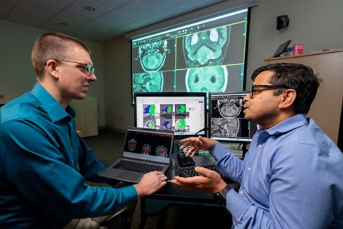
The laboratory employs advanced magnetic resonance imaging (MRI) techniques, such as diffusion-weighted imaging (DWI), perfusion-weighted imaging (PWI) and magnetic resonance spectroscopy (MRS) to achieve detailed characterization of diverse CNS tumors in pediatric patients. These methods facilitate the assessment of tumor cellularity, vascularity and metabolic profiles, enabling differentiation between benign and malignant lesions. Complementing these MRI approaches, we utilize novel positron emission tomography (PET) imaging modalities, including methionine (MET) PET and translocator protein (TSPO) PET to enhance diagnostic specificity and assessment of tumor microenvironments. Through the integration of the multimodal imaging data generated from these modalities, the laboratory ensures a comprehensive and accurate delineation of CNS tumors, supporting early and tailored therapeutic interventions.

AI-Based Techniques for Prediction of Tumor Types and Subtypes
To predict tumor types and subtypes with high precision, the laboratory leverages a variety of artificial intelligence (AI) models. These models are trained on large datasets derived from integrated multimodal imaging to classify tumors based on radiological patterns, texture analysis and quantitative biomarkers. Our multifaceted AI approach improves diagnostic accuracy and enables prognostic modeling, such as predicting molecular subtypes (e.g., different molecular types of medulloblastomas), which facilitates personalized treatment strategies.
Multimodal Imaging for Quantitative Estimation of Therapy-Associated Damage to Normal Brain Tissues
The laboratory utilizes advanced MRI techniques, including functional MRI (fMRI), diffusion tensor imaging (DTI) and quantitative susceptibility mapping (QSM) alongside novel TSPO PET imaging to quantitatively assess therapy-induced damage to normal brain tissues in pediatric neuro-oncology patients. These modalities enable the detection of subtle changes such as white matter integrity disruption, axonal injury and microstructural alterations resulting from chemotherapy, radiation, or surgical interventions. This quantitative assessment, along with changes in cognition profiles, will contribute to optimizing survivorship care, minimizing long-term neurocognitive deficits and enhancing overall quality of life for young cancer survivors.

Publications
Contact us
Asim Bag, MD
Associate Member, St. Jude Faculty
Director, Neuroimaging Section
Department of Radiology
MS 220, Room I3106
St. Jude Children's Research Hospital

Memphis, TN, 38105-3678 USA GET DIRECTIONS