St. Jude Family of Websites
Explore our cutting edge research, world-class patient care, career opportunities and more.
St. Jude Children's Research Hospital Home

- Fundraising
St. Jude Family of Websites
Explore our cutting edge research, world-class patient care, career opportunities and more.
St. Jude Children's Research Hospital Home
- Fundraising
Technology advances unlock the mystery of mitochondrial DNA — our “other” genome
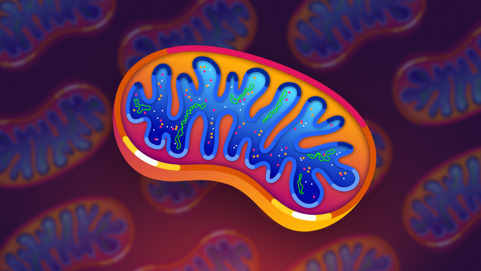
Mitochondria have their own genome. With advancements in analytical technologies, scientists at St. Jude Children’s Research Hospital are uncovering how this stowaway DNA can affect disease progression, including cancer.
About 1.8 billion years ago, one of our earliest ancestral cells engulfed a bacterial cell. Rather than being digested, the bacterium survived, and the two cells formed a partnership that persists to this day. Over time, that bacterium evolved into mitochondria, organelles found within cells whose role is echoed in science classrooms as being “the powerhouses of the cell.”
While most organelles, such as the nucleus, have functions that align closely with their evolutionary history, mitochondria retain unique quirks from their bacterial ancestry. Most notably, even though much of the original bacterial genome has been lost or transferred to the nucleus, mitochondria still harbor remnants of their original DNA.
Mitochondrial DNA (mtDNA) contains just 37 genes. Most of these genes are tied to the organelle’s role in the process of oxidative phosphorylation. This process converts nutrients from food into ATP, the cell’s primary energy currency. Beyond energy production, mtDNA has been used to trace human migration and lineage, solve historical mysteries, and even help restore Beethoven’s music. Yet, for decades, its role in disease, especially cancer, has been largely overlooked, as research has focused overwhelmingly on mutations in nuclear DNA. In cancer biology, the nuclear genome has long taken center stage, leaving the contributions of mtDNA as a relative afterthought. Now, St. Jude researchers are revealing how this genomic remnant might have a bigger impact than previously thought.
Mondira Kundu, MD, PhD, Department of Cell & Molecular Biology, investigates how mutations in mtDNA influence cancer, particularly acute lymphoblastic leukemia (ALL). While the stowaway DNA in mitochondria has fascinated researchers since its discovery in 1963, new technologies now put Kundu in a strong position to uncover how the cell’s “other” genome contributes to cancer.
A puzzle within a puzzle
“The first mitochondrial DNA mutations in cancer were confirmed when researchers saw certain tumor cell lines grow faster with specific mutations, but it wasn’t clear if the mutations actually caused this effect,” Kundu said. “Early studies catalogued mitochondrial DNA mutations in various cancers, but sample sizes were small, and findings were inconsistent.”

Through collaborative efforts spanning single-cell RNA sequencing and computational analysis, Mondira Kundu, MD, PhD, St. Jude Department of Cell & Molecular Biology (left), pictured with Kelly McCastlain, MPH, a scientist the Kundu laboratory, investigates how mutations found in mitochondrial DNA (mtDNA) contribute to tumor development.
Studying mtDNA presents unique challenges. Unlike DNA found in the nucleus, where mutations are copied when cells replicate, making interpretation straightforward, each cell contains hundreds or thousands of replicating mitochondria, each with its own mtDNA. This means a mutation may be present in only a fraction of a cell’s mitochondria, complicating efforts to assess its impact.
Advanced sequencing technologies have helped researchers identify links between mtDNA mutations and cancer. Still, Kundu cautions that not all mutations are created equal.
“Just because a mutation is predicted to be harmful doesn’t mean it actually drives disease in the body,” she explained. “Many mutations are simply passengers with no real impact, while only a few are true drivers that actively promote cancer development.”
Driver mutations are usually positively selected, meaning they are found at high frequencies in cancer cells. In contrast, most mtDNA mutations are present at relatively low levels when analyzing bulk tumor samples. At first glance, this low percentage might suggest that these mutations are not contributing to tumor development or progression. However, simply looking at the overall percentage fails to capture how mtDNA mutations are distributed within individual cells.
As Kundu explained, “If 20% of the total mitochondrial DNA is mutated, it could mean that 20% of cells have all of their mitochondrial DNA mutated, or that every cell has only 20% of its mitochondrial DNA mutated. These scenarios are very different, and understanding which is true is important for knowing how the mutation might impact the disease.”
mtDNA mutational ”sweet-spot” exacerbates cancer
In light of this, a 2025 Science Advances publication revealed just how significant a low-to-moderate fraction of mutated mtDNA can be. In this study, Kundu’s team found that while cells with a high fraction of mutated mtDNA showed suppressed growth, those with moderate levels experienced accelerated growth — a “sweet spot” where cancer thrives.
“We hypothesize that moderate metabolic stress may make cells more susceptible to transformation by different oncogenes,” Kundu said. “While the fully mutated state essentially shuts down potential, the moderately mutated state may open up cancer growth possibilities, even more than when there are no mutations.”
Having uncovered this phenomenon in mouse models, the team next wanted to determine whether the same pattern held true in human cancer samples. To do this, they needed to answer a critical question: When examining a low-to-moderate fraction of mutated mtDNA, is it 20% of mitochondria in 100 cells, or 100% of mitochondria in 20 cells? To develop the tools necessary to tackle this challenge, Kundu needed to find the correct team.
Looking at mtDNA one cell at a time
For the second study, also published in 2025 in Science Advances, Kundu recruited Gang Wu, PhD, Center for Applied Bioinformatics director and Catherine Welsh, PhD, formerly of the Department of Mathematics & Computer Science at Rhodes College, currently of Hope College, to help develop pipelines to identify tumor-specific mtDNA mutations from bulk sequencing data from the St. Jude–Washington University Pediatric Cancer Genome Project and to measure the mtDNA mutation load in individual cells using RNA sequencing data. She also brought in Stanley Pounds, PhD, Department of Biostatistics, who developed Mitovolve, a statistical model to distinguish between neutral drift and mutation selection.
The team used these tools to analyze the Kundu Lab’s single-cell RNA sequencing data to classify mtDNA mutations in terms of their distribution pattern and selection. By linking nuclear and mtDNA data, the researchers were also able to see if certain mtDNA mutations matched specific groups of tumor cells.
“If the mutated fraction is higher in certain subclones, that’s the key to understanding its role,” Wu said.
A breakthrough in connecting mitochondrial genetics to functional outcomes came from a collaboration with Jiyang Yu, PhD, Department of Computational Biology. Yu’s lab had previously created Network-Based Integrative Analysis (NETBID), a powerful computational approach for identifying key drivers of disease and therapy response.
By applying NETBID to the single-cell data, the collaborators pinpointed molecular networks that link mtDNA mutations to pathways involved in how cells grow and how leukemic cells respond to therapy. This analysis uncovered driver genes and pathways that are activated or suppressed in cells with specific mitochondrial mutation patterns, providing a potential link between mitochondrial genetics and functional outcomes.
Tools to tackle many mitochondrial diseases
Collectively, these tools enabled the team to determine whether mutations were harmless passengers or drivers that help cancer grow. However, to begin addressing individual mtDNA mutations’ contribution to disease requires selectively introducing point mutations which mimic those seen in cancer — a labor-intensive task due to the unique nature of mtDNA.
In a 2025 Computational and Structural Biotechnology Journal study, however, Kundu and Wu introduced MitoEdit, software that streamlines this process. “Many groups, including our own, have started exploring mitochondrial DNA engineering,” Kundu said. “MitoEdit makes it easier to identify target sites for editing. This program helps us generate constructs for editing mitochondrial DNA with less trial and error.”
These advancements reach well beyond cancer, because mitochondria can contribute to many diseases. St. Jude is meeting the challenge of understanding these diseases with the recent investment in Neurometabolic Translational Research, a component of the Pediatric Translational Neuroscience Initiative (PTNI) directed by Andrea Gropman, MD.
“For secondary mitochondrial disfunctions, such as cancer, we’re trying to see the ripple effect: If you change one cellular component, how does that ripple across systems?” Gropman said. “But with the ability to sequence both the nuclear genome, which includes mitochondrial genes, and mitochondrial DNA, we’re also identifying almost weekly new primary disorders, which present as stroke-like episodes, muscle disease or neuropathy.”
As technologies advance, the mysteries of mtDNA are coming into focus. No longer a footnote in pathology, mutations to the ancient bacterial DNA are now recognized as key players in disease development. These advancements are not only reshaping our understanding of disease but also paving the way for more targeted therapies, proving that the cell’s “other” genome is more than the sum of its parts.
About the author