St. Jude Family of Websites
Explore our cutting edge research, world-class patient care, career opportunities and more.
St. Jude Children's Research Hospital Home

- Fundraising
St. Jude Family of Websites
Explore our cutting edge research, world-class patient care, career opportunities and more.
St. Jude Children's Research Hospital Home

- Fundraising
David Solecki Lab
Understanding the link between cell polarity signaling and neuronal differentiation during cerebellar development.
About the Solecki Lab
Immature nerve cells must move or migrate in the developing brain to form connections called neuronal circuits. As these nerve cells move, they also mature. This maturation process, called differentiation, is an essential part of neurons migrating and linking up with other neurons inside a brain circuit. By examining how these neurons mature and differentiate, our laboratory strives to understand how nerve cells build the brain during fetal or early postnatal development. Our work focuses on the impact of improper neuron differentiation and how it can lead to neuronal defects and pediatric tumorigenesis.

Our research summary
The work of our laboratory sits at the interface of developmental neurobiology and technology development to produce a quantitative and mechanistic entry point to understand cell polarization and neuronal differentiation processes. We work with a strong team mindset to examine the differentiation process to discern the role of cell polarity pathways in disease and neuronal disorder formation. To propel our field of study forward, we also apply a collaborative spirit to help create the advanced technology needed to conduct our research.
The role of neuronal polarity pathways in development and disease
A major project that touches all areas of our laboratory is our investigation of neuronal polarity pathways. Our interest in understanding the basic development of the brain and the origins of disease spurs our research into this vital developmental pathway. Everything that happens in a differentiating neuron—how it generates cellular asymmetry and decides its cellular fate—relates to acquiring these specialized signaling pathways. Because these polarity signaling pathways are necessary for a neuron to differentiate, we work to identify the pathways' upstream regulators and downstream effectors.
Our recent research shows oxygen tension is an upstream regulator that manages the onset of neuronal polarity and normal differentiation. In terms of downstream effectors, our interest focuses on how polarity signaling regulates chromatin organization in the nucleus and cytoskeletal-adhesion coordination in the cytoplasm for neurons to migrate when they reach the differentiation process. Understanding these aspects of polarity signaling pathways sheds light on how these pathways impact development and disease formation.
Technology development
To ask the questions we ask, we need to break barriers in terms of what technology allows us to do and how it enables us to answer these questions. Sophisticated microscopes help us make detailed videos of maturing neurons in a healthy or diseased state, and we then use computational techniques, like image analytics or virtual reality, to determine the genes required for these processes.
To make certain aspects of biology more visible, our initial work focused on developing fluorescent protein probes to show where sites of intercellular contact recruit cell adhesion molecules. Our work with these probes now centers on intersectional fluorescent protein probes that can detect multiple epigenetic marks in live cells without the need for antibodies.

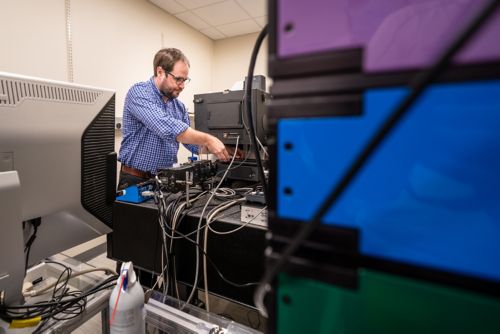
We also work in collaborative efforts to advance the capabilities of microscope technology. One exciting endeavor is our work with the lattice light sheet microscope (LLSM), which can image cells rapidly with isotropic resolution and little phototoxicity. We were the first in the country to adopt a commercial version of this microscope (2016), and such access permitted us to publish the first two papers in neurobiology to use LLSM. Our work with LLSM allows us to see how live neurons rapidly repack the DNA in their nucleus as they express new genes during differentiation. Our access to this type of technology improves our capabilities in microscopy and imagery data analysis since the microscope can produce 2GB of data per second.
Through our work with LLSM, we forged a collaboration with Janelia Research Campus, including Erik Betzig and Harald Hess, where we examine chromatin organization for the first time with incredible detail. This collaboration centers around a dual-microscopy approach called cryo-SR/EM microscopy. This approach images high-pressure frozen cells—preserved in their most life-like form—with super-resolution light microscopy and an electron microscope. Using computational techniques, we layer the vivid image of cellular machinery—captured via light microscopy—over the black-and-white images captured by electron microscopy. These layered cryo-SR/EM images, taken before and after differentiation, help identify the nuclear domains that change in the differentiation process and reveal that neurons pack and/or unpack unique classes of genes. This information is vital in our understanding of the differentiation process and how neurons organize gene expression.
The multidisciplinary approach of our lab, coupled with our commitment to technological advancement, allows us to search for answers to some of developmental neurobiology’s most pressing questions. Our commitment enables us to provide insights into the developing brain that help us understand the origins of neuronal disease and disorders in children.
Selected publications
Contact us
David Solecki, PhD
Member
Developmental Neurobiology
Director, Division of Neuronal Cell Biology
MS 323, Room M2412
St. Jude Children's Research Hospital

Memphis, TN, 38105-3678 USA GET DIRECTIONS