St. Jude Family of Websites
Explore our cutting edge research, world-class patient care, career opportunities and more.
St. Jude Children's Research Hospital Home

- Fundraising
St. Jude Family of Websites
Explore our cutting edge research, world-class patient care, career opportunities and more.
St. Jude Children's Research Hospital Home

- Fundraising
Imaging provides a front row seat to metabolic reactions

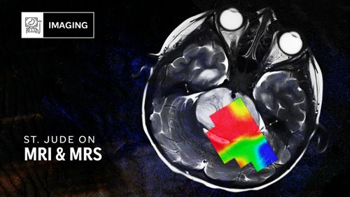
Magnetic resonance imaging (MRI) is a valuable tool that allows researchers to see structures inside the body, even making it possible to study metabolic processes.
Within living beings, a metabolic opera is being produced. The stars of the show are molecules performing various functions. The stage is set within the body, and the villain lurks in the shadows: a disease such as cancer. To triumph over this formidable foe, researchers must acquire a profound understanding of the intricacies of a healthy body and the biological elements that propel cellular dysfunction. Armed with this knowledge, they can devise therapeutic strategies aimed at curing or, at the very least, slowing down the disease.
At St. Jude, I lead the metabolic imaging research lab, where we identify, explore and establish clinically feasible non-invasive imaging approaches. Our lab offers the ability to glimpse into the intricate world of biochemicals and metabolic processes that power our bodies.
Our primary focus is unraveling the pathways involved in disease. These pathways are a linked series of chemical reactions that occur within a cell. To do so, our team unleashes an arsenal of imaging techniques, relying on the formidable powers of molecular magnetic resonance imaging (MRI) and spectroscopy (MRS). Through this work, we endeavor to develop clinically translatable methods that can significantly impact patient outcomes.
Unveiling the metabolic odyssey
Our body resembles an expansive ocean of water molecules. Some water molecules freely circulate, while others are bound within cells to membranes, proteins and macromolecules. Within the confines of an MRI scanner and its potent magnetic field, the protons in water align themselves with this force. Introducing a perpendicular magnetic field, called a radiofrequency pulse, gracefully tips over these protons. As the particles return to their original alignment, a minuscule current is generated by each one and recorded, capturing the nuclear magnetic resonance signals that were once concealed. This is how an MRI works to capture images.
This fundamental principle is not limited to water molecules. It extends to small molecules or metabolites too. In the imaging results, each peak intricately maps the presence of metabolites such as lactate, 2-hydroxyglutarate, phosphocholines, and others discreetly residing within the tissue. This mapping enables our lab to study specific metabolic pathways associated with a disease.
Metabolic insights into DIPG
Within my lab, we are deeply engaged in a project aimed at unraveling the metabolic vulnerabilities of diffuse intrinsic pontine glioma (DIPG). This aggressive brain tumor intricately weaves its way through vital structures such as the brainstem and pons, rendering biopsies impossible. Given that radiation therapy is the only currently available therapy, the prognosis for this cancer remains disheartening.
To better understand DIPG, we need to understand its metabolic properties. We use patient-derived samples in mouse models to meticulously track the tumor’s metabolism as the disease progresses. Our toolkit includes using chemical exchange saturation transfer (CEST) MRI to map metabolites such as lactate, glucose and free amines, enabling us to evaluate the dynamic response to targeted therapy noninvasively.
Furthermore, we delve into the world of metabolic pathways, monitoring flux rates to unveil hidden vulnerabilities within these tumors. While we can learn much from injecting trackable, or “tagged” metabolites into our models, doing so demands additional expertise and specialized imaging coils. Even then, results may not match the spectral resolution needed.
Our lab has created new groundbreaking techniques for quantitative exchanged-label turnover MRS (qMRS), which quantifies the dynamics of metabolism to address this challenge. Through this process, we can offer a new avenue in MRS research that provides valuable insights into the metabolic intricacies of DIPG.
Searching for metabolic biomarkers
In addition to DIPG, our team is also seeking to apply metabolic imaging techniques to better understand Ewing sarcoma, a rare bone or soft tissue cancer prevalent among children and teenagers. Ewing sarcoma is characterized by a pivotal gene, SLFN11, which can be used to determine the likely success or failure of chemotherapy. However, SLFN11 isn’t perfect in this regard, suggesting that intricate metabolic changes underpin Ewing sarcoma’s aggressive growth and influence treatment response. We aim to uncover metabolic biomarkers closely linked to SLFN11 using cutting-edge technologies such as transcriptomics, metabolomics, lipidomics and metabolic imaging. This pursuit holds the promise of unveiling novel therapeutic strategies.
We are also actively searching for metabolic biomarkers to better understand the intricacies of cancer treatment-associated cognitive decline and frailty in survivors of childhood cancers. A profound comprehension of these mechanisms is imperative for the development of targeted therapies in the future. Research from St. Jude investigators, among others, suggests that childhood cancer survivors seem to be on an accelerated aging trajectory. Developing non-invasive, sensitive and reliable metabolic imaging methods to monitor survivors over time is paramount to unraveling connections between accelerated aging and metabolic signaling. Biomarkers will play a pivotal role in guiding future therapy design and optimizing intervention dosages.
Embarking on muscle exploration with CrCEST
Have you ever stopped to consider how our muscles work? What can seem like magic is actually a fundamental biologic process where the key to energy — mitochondrial oxidative phosphorylation (mtOXPHOS) — fuels our every move. While phosphorous (31P)-MRS has long been the champion and gold standard for mtOXPHOS assessment, its limitations open the door to the potential alternative of creatine-weighted CEST (CrCEST) MRI.
CrCEST can decipher the post-exercise recovery rate, a task that demands precision in delineating muscle boundaries. Our findings from leg muscle exercises showcase the CrCEST technique's commendable repeatability (across multiple experiments on the same subject) and reproducibility (consistency across different readers interpreting the same experiment), especially during plantar flexion exercises.
Our laboratory is exploring the application of CrCEST to study neuromuscular disorders. The potential of CrCEST in diagnosing muscle health positions it as a promising tool for diverse populations grappling with muscle disorders or mitochondrial diseases.
The power of imaging for metabolic research
In our lab, collaboration is the cornerstone of our pursuit to unravel the cellular-level mechanisms propelling various cancers in pediatric patients using MRI. Our team is a dynamic tapestry of expertise, bringing together researchers from diverse disciplines such as biophysics, biochemistry, cell biology, computation and MRI engineering.
This interdisciplinary approach enables us to gain profound insights into the intricate workings of the cell. Together, we are actively developing robust pipelines for clinically feasible cutting-edge imaging tools. Our mission extends beyond merely understanding the role of metabolism in cancer development; we are equally committed to shedding light on aspects of cancer survivorship and exploring connections with neurodevelopmental disorders.
If metabolic processes are an opera playing out in the body, we aim to be the stage crew — utilizing all the tools at our disposal to understand how the show runs and how to deal with it when things go awry. MRI and MRS are two of our most valuable tools, giving us a behind-the-scenes look at metabolic processes that can inform how cancer and other diseases are treated.


