St. Jude Family of Websites
Explore our cutting edge research, world-class patient care, career opportunities and more.
St. Jude Children's Research Hospital Home

- Fundraising
St. Jude Family of Websites
Explore our cutting edge research, world-class patient care, career opportunities and more.
St. Jude Children's Research Hospital Home

- Fundraising
Scientists are knowledge hunters who seek an objective understanding of the world. Acquiring this understanding relies on the tools a scientist has in their toolkit. Light is a critical tool.
It illuminates the material world and can reveal what is hidden by darkness. St. Jude researchers work on the cutting edge of science, often butting up against physical boundaries that limit the types of biological questions they can answer. However, researchers have shown that they can push past the boundaries of what has been possible, creating new approaches to peer into the unknown. The resulting discoveries illuminate science, lighting the way to knowledge.

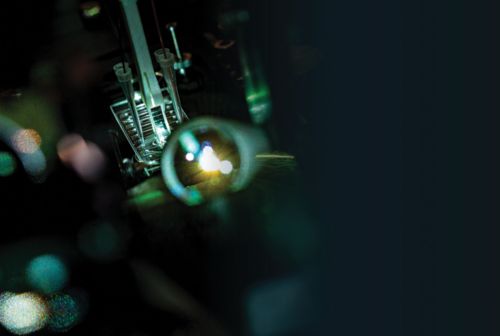
Total internal reflection fluorescence (TIRF) microscopy is a powerful imaging technique that relies on a prism to direct light into the chamber in such a way that only the sample is illuminated — a concept called optical sectioning.
Go ahead, FRET the small stuff
Fluorophores are molecules that can absorb visible light, which in turn enables electrons to jump to higher energy states. Upon relaxation of the electron, that energy is sometimes released as glowing light (fluorescence). Combining fluorescence with microscopy is a powerhouse pairing that can detect cellular structures and biological processes, which can inform foundational biology and the diagnosis and treatment of human diseases.
Biomolecules change their shape, or conformation, to interact with binding partners and complete physiological functions. These movements can be subtle and localized — occurring on a time scale around one quadrillionth of a second — or more exaggerated, on a time scale of microseconds to seconds. Understanding how biomolecules move while they work is key to understanding their function.

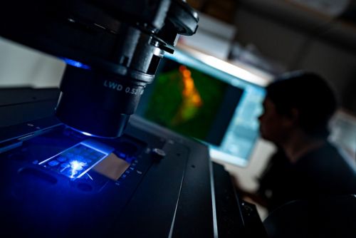
A postdoctoral researcher uses fluorescence microscopy to study cellular diversity.
Technological advances in microscope mechanics, improved data processing, and reimagined fluorophores have increased the speed and precision of fluorescence microscopy. Scientists can now study the movements of individual molecules in real time by using single-molecule Förster resonance energy transfer (smFRET). This method works by placing two or more fluorophores on different parts of a molecule, exciting the sample with light, and measuring the energetic communication among the fluorophores. Quantifying the energy transfer between fluorophores informs scientists about the distance between them, which can be on the scale of one-billionth of an inch. This level of sensitivity makes this technique ideal for measuring biological processes at the molecular scale.
One limitation of fluorescence-based imaging methods is that fluorophores can fail to fluoresce because of a change in a fundamental property of the excited electron called its spin state. “Every time an electron is excited, there’s a probability that it will lose memory of its spin and adopt an inverted spin state,” said Scott Blanchard, PhD, Department of Structural Biology. “If this occurs, it cannot relax back to its ground state and becomes trapped in a highly reactive, nonfluorescent triplet state that is 100,000 times longer-lived.”
Triplet states, called “dark states,” impact a fluorophore’s brightness and durability and cause phototoxicity. Dark states also reduce data quality and accuracy.

What the field believed was that triplet states were an immutable reality. But we had no other choice but to try to find solutions.
Department of Structural Biology
Blanchard surmised, “What the field believed was that triplet states were an immutable reality. But we had no other choice but to try to find solutions.”
The need to succeed led Blanchard’s team to intramolecularly tether triplet state quenchers to fluorophores. This discovery gave researchers a generalizable strategy to reduce dark state lifetimes in fluorophores by as much as 1000-fold. Fluorophores imbued with this property are called “self-healing.”
Published in Nature Methods, this study by Blanchard’s team demonstrated for the first time that self-healing fluorophores enable more robust and precise molecular-scale measurements. This transformative work substantially advances smFRET imaging and helps ascertain the function of biological molecules. However, the self-healing technology has the potential to benefit all forms of fluorescence imaging, which Blanchard hopes will be advantageous to fluorescence microscopy pursuits at St. Jude and around the world.

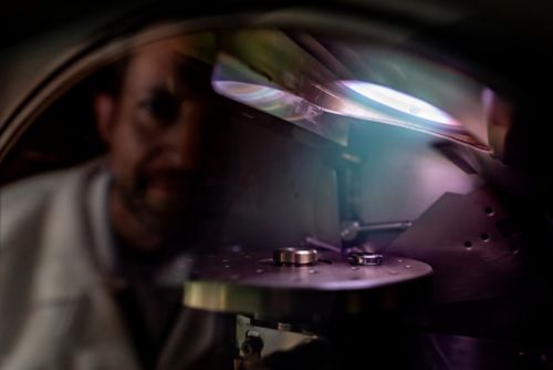
Cam Robinson, PhD, Cell and Tissue Imaging Center, performs metal sputter coating of a sample to increase its conductivity before imaging on the scanning electron microscope.
Viewing light through an intersectional lens
Necessity is the mother of invention, and Lindsay Schwarz, PhD, Department of Developmental Neurobiology, agrees with that ancient Greek proverb. Schwarz researches norepinephrine-expressing (NE) neurons, which represent about 0.00003% of brain cells. These cells modulate anxiety, attention, memory, and stress responses.
Aberrant responses by NE neurons can cause mental health issues, including anxiety and depression. “We treat anxiety and depression with drugs that target norepinephrine signaling, but these drugs act globally, meaning there’s often a detriment to other important functions for norepinephrine that you don’t want to see,” explained Schwarz. “Targeting these neurons more specifically could help ameliorate that.”
As Schwarz postulated how to target subsets of neuronal cells, she realized that she needed a way to isolate subpopulations expressing overlapping elements or genetic features, a user-friendly intersectional approach — something that did not exist.
Writing in Nature Neuroscience, Schwarz and former St. Jude graduate student Alex Hughes detailed the development of Conditional Viral Expression by Ribozyme Guided Degradation (ConVERGD). ConVERGD is an intersectional expression platform pairing adeno-associated virus vectors, which insert genetic material called transgenes, with ribozymes (RNA strands that catalyze reactions) to express desired traits in specific cellular subsets.
ConVERGD constructs activate transgenes and fluoresce only when two protein recombinases, Flp and Cre, are present. Flp ensures the transgene is read properly, and Cre abolishes ribozyme activity that would otherwise cleave the transcribed mRNA and prevent protein production. Flp and Cre can be introduced into many cells and organisms and can be activated independently through functional programs that are distinct to specific cells. Utilizing these molecular elements in the way that ConVERGD does allows specificity in identifying cell lineages and subsets in living organisms based on multiple parameters, highlighting the versatility of this approach. ConVERGD can also be used in other systems beyond the brain.

These methods are now being used by other labs to study important processes in the brain in ways that haven’t been done before.
Department of Developmental Neurobiology
As proof of concept, Schwarz’s team looked for NE neurons that also expressed prodynorphin (Pdyn), which was thought to be involved in anxiety. Using ConVERGD, the researchers fluorescently labeled a targeted subpopulation of neurons expressing both Pdyn and Dbh, which makes norepinephrine. Further behavioral studies linkedthis subpopulation to a specific behavioral response.
ConVERGD-based viral tools made it possible for Schwarz’s team to identify the anatomical connectivity and behavioral influence of Pdyn+/Dbh+ NE neurons and helped the field understand how this unique population of neurons provokes such diverse behavioral states — a question that has fascinated researchers for years.
Schwarz continues to expand the ConVERGD toolkit. She said, “These methods are now being used by other labs to study important processes in the brain in ways that haven’t been done before.”
Seeing is believing
How researchers experimentally use illumination varies as technology advances. This is the case in the laboratory of Stacey Ogden, PhD, Department of Cell & Molecular Biology, who studies the role of signaling molecules such as Sonic Hedgehog (SHH).
Precisely when and where a cell receives an SHH signal is tightly regulated during development to ensure tissues and organs pattern properly. Still, scientists were unclear about how SHH reaches target cells with appropriate precision. Some wondered if thread-like cellular projections called cytonemes were involved, but no one had visualized the structures in mammalian tissue — not until Ogden reevaluated the capabilities of imaging science and, quite literally, saw cytonemes in a new light.

For a long time, visualizing these structures in complex developing mammalian tissue has been challenging, but we’ve finally found a way.
Department of Cell & Molecular Biology
“For a long time, visualizing these structures in complex developing mammalian tissue has been challenging,” Ogden said. “But we’ve finally found a way.”
In Cell, Ogden described combining modern microscopy with optimized sample preparation to see fluorescent thread-like protrusions extending from cells. Ogden’s team systematically tested various methods of sample preparation, fixation, and imaging to ascertain the most conducive protocols for imaging in mammalian tissue.

A St. Jude scientist prepares tissue samples for scanning electron microscopy
To effectively visualize the cellular membrane extensions, the team leveraged a genetic lineage tracer in which membranes of SHH-expressing cells are labeled with a fluorescent protein. To preserve the 3D structure, tissues expressing the fluorescent marker were sectioned with a device called a vibratome, which allowed the researchers to create tissue sections that were thick enough to preserve cytonemes but thin enough to facilitate imaging of the membrane marker. Sectioning with the vibratome also minimized the fracturing of cytonemes that typically occurs when sectioning tissue with a more standard device called a cryostat.
Vibratome sections were visualized using a confocal microscope that combines super-resolution imaging with the ability to track fluorescence lifetime. This approach allowed the researchers to obtain images with lower background noise and higher spatial resolution. Ogden’s team also used scanning electron microscopy for additional validation that what they observed using light microscopy was occurring at a larger scale.
The researchers were able to observe the tip-to-tip connections made by these cellular extensions in the neural tube, a structure found early in development that eventually becomes the brain and spinal cord. These tip-to-tip connections appeared to join cells at the apex of the membranes lining the lumen (hollow space within the neural tube) to primary cilia at the apex of adjacent cells.
Comprehensively, Ogden’s work showed that cytonemes are key contributors to SHH deployment and rapid delivery for tissue patterning. Furthermore, the work showed that when cytoneme function was disrupted, aberrant SHH signaling ensued.
“This is the first demonstration of these cytoneme-based transport processes occurring during the development of a complex mammalian tissue such as the neural tube,” Ogden said.
Lighting the way forward
Illuminating discoveries are often made by pushing past the boundaries of what is known or what already exists to gain new understanding. This mindset of exploration is what enables researchers to gain fundamental biologic insights, whether improving smFRET, creating new tools such as ConVERGD, or seeing structures such as mammalian cytonemes for the first time. By lighting the way forward, these St. Jude researchers are forging the next generation of discoveries.