St. Jude Family of Websites
Explore our cutting edge research, world-class patient care, career opportunities and more.
St. Jude Children's Research Hospital Home

- Fundraising
St. Jude Family of Websites
Explore our cutting edge research, world-class patient care, career opportunities and more.
St. Jude Children's Research Hospital Home
- Fundraising
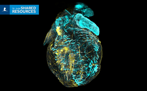
Cell and Tissue Imaging Center: Electron Microscopy
Accelerating the use of ultrastructural imaging technologies across St Jude
OVERVIEW
The Cell and Tissue Imaging Center is a fully staffed microscopy core available to all St. Jude researchers. Its mission is to drive and accelerate the use of a broad spectrum of sophisticated light microscopy, electron microscopy and image data processing technologies. Expert imaging scientists are on hand to guide and assist throughout the experimental process up to the point of publication. As well as instrument training and operation, helping with sample preparation, and guiding analyses, staff can help to select the best imaging strategies to answer specific research questions. The Light Microscopy and Electron Microscopy divisions also collaborate to provide correlated light and electron microscopy imaging. Costs are heavily subsidized by the institution to speed discovery.


IMPACT
The CTIC-EM is a highly specialized resource providing 2D and 3D nanometer scale (1/100,000 the width of a human hair) imaging of cells and tissues to St. Jude researchers. The facility houses an array of leading-edge imaging systems and a staff with expertise in multiple areas of ultrastructural imaging including immunogold labeling, Tokuyasu cryoultramicrotomy, and volume EM. Staff are available for individual consultations on project set-up, optimization, and initiation and guidance on image analysis as requested and in conjunction with the Center for Bioimage Informatics.
By the numbers
Ranked as an ‘Exceptional’ shared resource by the 2025 Cancer Center external review panel
By the numbers
15
Collaborations with over 15 labs
By the numbers
30
On average, the CTIC-EM is acknowledged on about 30 publications annually
By the numbers
700
The facility processes an average of 700 samples for electron microscopy annually.
In the news
Shared Resource Spotlight: Cell and Tissue Imaging Center
The Cell and Tissue Imaging Center helps fuel breakthroughs using light and electron microscopy.
EQUIPMENT / SERVICES
Instrumentation
Thermo Scientific Helios Laser Hydra
multi-gas plasma focused ion beam scanning electron microscope providing large area subcellular 3-dimensional imaging with a choice of plasma sources to suit sample needs and a femtosecond laser to speed sample preparation
Thermo Scientific Tecnai G2 F20-Twin
transmission electron microscope equipped for electron tomography and providing resolution to 0.27 nm
Zeiss GeminSEM 460
field emission scanning electron microscope equipped for high speed, large area imaging of samples as well as variable pressure operation for non-conductive samples
Zeiss Crossbeam 550
focused ion beam scanning electron microscope providing 3-dimensional imaging as sub-cellular resolutions and a gallium ion beam for sample milling.
Preparatory equipment
(1) Leica EM ICE High Pressure Freezer
(2) Leica AFS2/FSP systems for processing frozen samples
(2) Leica EMTP systems for room temperture processing
Pella BioWave microwave
(1) Tousimis AutoSamdri critical point drier
(4) Leica UC7/ARTOS ultramicrotome systems with cryoultramicrotomy capabilities
Denton and Leica high vacuum coating systems
About the Director
-
View Details
Camenzind Robinson, PhD
Director, Electron Microscopy, Cell and Tissue Imaging Center
Email
cam.robinson@stjude.orgCamenzind Robinson, PhD, earned his PhD in Cell Biology from Yale University and has more than 20 years of experience utilizing electron microscopy to answer key biological questions. Before joining St. Jude in 2018, Dr. Robinson has provided EM support to individual Howard Hughes Medical Institute laboratories at Duke University and at HHMI’s Janelia Research Campus. Robinson has also led centralized and shared resources at the US Army Research Institute of Infectious Diseases. His expertise is in sample preparation and transmission EM, with proficiency in scanning EM, immunogold labeling, negative staining and volume EM.
Meet the Team
-
View Details
Woo Jung Cho, MSc
Senior Scientist
-
View Details
Randall Wakefield
Scientist
-
View Details
Nathalie Becerra Mora, PhD
Associate Scientist
-
View Details
Amanda Johnson, MS
Senior Researcher
-
View Details
Sarah LaGrange
Researcher