St. Jude Family of Websites
Explore our cutting edge research, world-class patient care, career opportunities and more.
St. Jude Children's Research Hospital Home

- Fundraising
St. Jude Family of Websites
Explore our cutting edge research, world-class patient care, career opportunities and more.
St. Jude Children's Research Hospital Home

- Fundraising
T cells are the body’s frontline protectors, pivotal in the immune system’s defense against disease.
Understanding T-cell biology is of great significance for immunotherapy, in which genetically modified T cells have demonstrated potential to treat cancers once considered incurable. However, while these therapies have offered a chance at remission to some patients, they have not been universally successful.
The challenge of immunotherapy lies in optimizing T-cell performance — enhancing their activation, longevity, and function to ensure they respond robustly and sustainably. Understanding these processes is central to improving immunotherapies as researchers explore ways to “fine-tune” T cells, thereby preventing their limitations and ensuring they function at their peak for longer periods.
At St. Jude, researchers harness the power of T cells by diving deep into their biology and discovering how to enhance their precision and persistence. These advances are opening the door to more effective therapies, bringing us closer to harnessing the power of the immune system to target and destroy tumors with minimal side effects.

Our research uncovers the mechanisms by which T cells adjust to extracellular nutrient conditions and connects these mechanisms to a key intracellular organelle, the lysosome.
Department of Immunology
Foundations of tissue-resident and regulatory T-cell function
To better understand and optimize the therapeutic application of T cells in cancer and other diseases, researchers at St. Jude use fundamental T-cell biology to unlock new ways to enhance immune efficacy. Hongbo Chi, PhD, Department of Immunology, studies metabolic systems and signaling pathways in T cells and capitalizes on those pathways to harness T cells’ antitumor and tissue immunity functions.
“T cells migrate to various tissues and must adapt to the local nutrient environment. Our research uncovers the mechanisms by which T cells adjust to extracellular nutrient conditions and connects these mechanisms to a key intracellular organelle, the lysosome,” Chi explained.

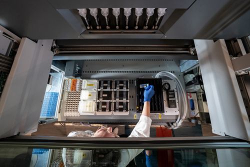
Scientists at the Hartwell Center for Biotechnology use state-of-the-art equipment to perform microarray analyses.
Tissue-resident memory (TRM) and TRM–like cells provide rapid, localized immune responses at the site of infection or tumor growth. Chi’s team used CRISPR-Cas9 genetic screens to uncover critical signaling pathways influencing TRM cell development and differentiation. They found that TRM cell formation depends on processes in cellular organelles called mitochondria. Additionally, signaling nodes at the lysosome, such as Folliculin (Flcn), Ragulator and Rag GTPases, restrict TRM formation and development.
Apart from organelle signaling, the study also found that nutrient availability played a role in tissue immunity. Specifically, Flcn modulates the activity of transcription factor EB (Tfeb). Flcn–Tfeb signaling, induced by amino acid deprivation, contributes to TRM cell development. The relationship links nutrient stress to cell fate decisions. Therefore, the Flcn–Tfeb axis is a regulatory pathway that coordinates immune responses to nutrient availability and lysosomal function.
These findings, published in Immunity, highlight the complex interplay between metabolism and T-cell function and offer new insights into how T cells can be optimized to improve immune responses in tissues.
T-cell function is governed by metabolism and the ability to distinguish self from non-self, a process overseen by regulatory T (TREG) cells. The protein Foxp3 serves as a gatekeeper, or master regulator, determining what the immune system recognizes as “self” to protect from attack. However, the exact mechanism behind this process has remained elusive.

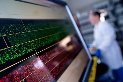
Microarray analysis allows the exploration of gene expression and epigenetic changes in T cells, offering valuable insights into their biology.
A study led by Yongqiang Feng, PhD, Department of Immunology, and published in the Journal of Experimental Medicine, examined Foxp3. For decades, Foxp3 has been considered a transcription factor, a protein that coordinates gene expression. The tunable nature and broad swath of responses that Foxp3 controls led Feng to question the biochemical nature of how this transcription factor was, itself, regulated. How did Foxp3 know whether to coordinate a suppressive immune response?

As immunologists, we don’t just want to understand the mechanisms; we want to know how we can take advantage of this knowledge to engineer better therapies.
Department of Immunology
The researchers studied the relationship between Foxp3’s protein-binding partners and its function, making a surprising discovery. “We found that when the environmental conditions changed, the ability of Foxp3 to interact with DNA also changed,” Feng said. “We found the Foxp3 does not directly interact very much with the DNA but rather binds to other DNA-binding proteins. In this sense, it is a transcriptional cofactor.”
These findings suggest that environmental triggers activating regulatory T cells drive the expression of Foxp3’s binding partners, which then coordinate with Foxp3 to establish the appropriate immune response. Foxp3 swaps out these binding partners depending on the environmental cue. These environmental triggers help Foxp3 regulate TREG cell function, control immune responses, and prevent autoimmune disease, but they can also promote tumor growth. This new paradigm proposes druggable strategies to modulate TREG cell function.
“By exploring the configuration of Foxp3 protein, we hope to identify new druggable targets and design a better protein, leading to better TREG cells, meaning better treatments,” said Feng. “As immunologists, we don’t just want to understand the mechanisms; we want to know how we can take advantage of this knowledge to engineer better therapies.”
In a separate study, Benjamin Youngblood, PhD, Department of Immunology, and Caitlin Zebley, MD, PhD, Department of Bone Marrow Transplantation & Cellular Therapy, in collaboration with researchers at the University of Minnesota, discovered that T cells can proliferate indefinitely without the typical functional decline seen in most cell types. This work, published in Nature Aging, revealed that T cells can outlive an organism, potentially enduring multiple lifetimes.

Tae Gun Kang, PhD, Department of Immunology, uses flow cytometry to research T-cell behavior and function.
To study this phenomenon, the researchers used specific biomarkers known as epigenetic markers that accumulate over time. This “epigenetic clock” tells a retrospective story about the life cycle of a cell independent of the organism itself. The accumulation of genetic mutations, the shortening of telomeres (the protective caps on chromosomes) and methylation patterns are currently regarded as the most accurate ways to interrogate the process of aging.
Through a collaboration with investigators at the University of Minnesota, the researchers utilized a mouse model that maintained the same line of T cells through several life cycles. Based on the markers of the epigenetic clock, the researchers found that the T cells were not bound by the reasonable limits of organism lifespan, surviving for up to four lifetimes. This study underscores the pivotal role of epigenetics in T-cell aging and function, revealing how molecular mechanisms extending beyond chronological age can influence cellular memory and longevity.

Our research efforts defining barriers limiting immunotherapy will guide our future efforts to engineer more effective treatments and grow our pediatric immuneoncology program.
Department of Immunology
Fine-tuning T cells to overcome immunotherapy challenges
Immunotherapies face challenges impacting their effectiveness, particularly maintaining functional persistence, which ultimately reduces the long-term efficacy of such treatments. When T cells are overstimulated, they become exhausted, a nonfunctional phase that significantly impairs their ability to destroy tumors. T-cell exhaustion has emerged as one of the primary reasons many T cell–based immunotherapies fail.
In a paper published in Nature Immunology, Youngblood and his colleagues identified the level of stimulation that leads to optimal anti-cancer performance. They found that preventing T-cell exhaustion requires precise stimulation. The study also revealed that how tightly a parental T cell binds to a cancer protein determines if its daughter cells will be anti-cancer effectors or exhausted. If binding strength is not just right, the progenitor T cells develop into exhausted cells.

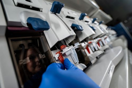
Advanced technology automates the loading of microarrays, enabling high-throughput analysis for cutting-edge research.
“We wanted to know how signal strength contributed to either the maintenance or the progression of T cells to a terminally exhausted state,” said Youngblood. “Understanding what controls the transition between progenitor to the dysfunctional exhausted state will allow us to develop engineering approaches that improve the longevity of T cell–based immunotherapies for solid and liquid tumors.”
Checkpoints are signals that tell T cells how to react to diseased cells or pathogens. Tumors can hijack these checkpoints to turn off the immune system. One type of immunotherapy, immune checkpoint inhibitors, blocks tumors’ ability to suppress T-cell function, thereby helping the immune system find and kill cancer cells.
Although the approach has shown remarkable success, it does not work for all patients. To understand why, Zebley and Youngblood collaborated with colleagues at the Van Andel Institute. The researchers explored what is different about the T cells of patients who respond to immune checkpoint inhibitors.

We showed Asxl1 disruption endows T cells with superior long-term therapeutic potential, which could be a promising strategy for the design of future T cell–based immunotherapies.
Department of Bone Marrow Transplantation & Cellular Therapy
“Looking at clinical trial data from myelodysplastic syndrome patients treated with checkpoint inhibitors, we saw that while most didn’t respond well, a small subset had long-term survival,” explained Zebley. “These patients had an ASXL1 mutation in the T cells, which led us to question whether the mutation could drive response to immune checkpoint inhibition.”
In a paper published in Science, the researchers reported that intentional Asxl1 disruption in murine T cells improves tumor control during immune checkpoint inhibition. The investigators also discovered that Asxl1 regulates the epigenetic checkpoint governing terminal T-cell differentiation into the exhausted state. Asxl1-depleted T cells resisted exhaustion for the animal’s lifespan and controlled a range of tumors, compared to T cells with Asxl1 intact, which become exhausted and control only a limited type of tumors.
“We showed Asxl1 disruption endows T cells with superior long-term therapeutic potential, which could be a promising strategy for the design of future T cell–based immunotherapies,” Zebley said.
Orchestrating breakthroughs in bone marrow transplantation
Breakthroughs in understanding fundamental T-cell biology pave the way for more precise and effective immunotherapies. “Support for fundamental science is one of the many things that makes St. Jude special,” explained Youngblood. “Our research efforts defining barriers limiting immunotherapy will guide our future efforts to engineer more effective treatments and grow our pediatric immune-oncology program.”
An example of how fundamental insights lead to tangible improvements is findings from a phase 2 clinical trial published in the Journal of Hematology & Oncology. The study, led by Brandon Triplett, MD, and Swati Naik, MBBS, both of the Department of Bone Marrow Transplantation & Cellular Therapy, shows promise in enhancing the donor immune attack on the host’s leukemic cells, called the graft-versus-leukemia effect, without causing excessive dangerous graft-versus-host disease (GVHD).
Patients with leukemia or other blood cancers who do not respond to chemotherapy often need hematopoietic cell transplantation, also called bone marrow or stem cell transplantation, to treat their cancer. Although potentially lifesaving, transplantation carries substantial risks. Finding a donor match is challenging, and using mismatched donors increases complications such as GVHD, which occurs when the transplanted T cells from the donor’s bone marrow identify the patient’s tissues as foreign and launch a harmful immune attack.

In this study, even without total body irradiation, the leukemia-free survival rates were comparable, if not better, than those seen in studies that used it.
Department of Bone Marrow Transplantation and Cellular Therapy
The researchers minimized this complication by selectively removing naïve T cells, which are defined as T cells that have not yet encountered the specific protein they were designed to recognize and which generally drive GVHD. By targeting only naïve T cells, the researchers could leave mature memory T cells behind. These memory T cells harness the beneficial graft-versus-leukemia effect and can protect patients against infections while minimizing the risk of GVHD.
“In bone marrow transplantation, total body irradiation is typically considered essential to achieve favorable outcomes,” explained Naik. “However, in this study, even without total body irradiation, the leukemia-free survival rates were comparable, if not better, than those seen in studies that used it.”

A scientist analyzes fluorescent signals on a microarray to gain insights into molecular activity in ongoing research.
“The thing about transplantation is that you’re building a whole new immune system in the recipient from the donor cells,” added Triplett. “We’re designing it to be as robust as possible against infections and leukemia while carefully minimizing the risk of graft-versus-host disease.”
As scientists deepen their understanding of T-cell biology and immune system activity, breakthroughs advance the development of fine-tuned and effective therapies. By enhancing the power of T cells and other immune components, St. Jude researchers are laying the foundation to improve immunotherapy treatment for patients with even the most intractable cancers.
We’re designing it (the immune system) to be as robust as possible against infections and leukemia while carefully minimizing the risk of graft-versus-host disease.
Department of Bone Marrow Transplantation and Cellular Therapy